1. In vivo kinematic analysis of total knee arthroplasty (TKA)
Total knee arthroplasty is one of the successful surgical procedures for patients with osteoarthritis of the knee. Several types of implants have been developed. However, it has been difficult to evaluate whether they function as designed in concept. We have developed a novel 2D/3D registration technique that estimates the in vivo spatial position (3D) from X-ray fluoroscopic images (2D). To improve patient’s satisfaction, we have analyzed and developed several implants.
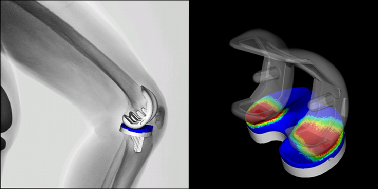
Fluoroscopic image during squatting (Left). A 2D/3D registration technique exhibits the spatial positon of the metallic components and polyethylene insert (Right).
2. Real-time 3D dynamic analysis in upper extremities
We have developed a technology -2D-3D Registration method, in collaboration with Nara Institute of Science and Technology (Prof. Sato and Assoc. Prof. Otake). This technique enables real-time 3D dynamic analysis by superimposing 3D bone models generated from CT data onto continuous X-ray fluoroscopic images. We have conducted real-time dynamic analysis of healthy forearm and of malunited forearm diaphyseal fractures, and published the results. Detailed dynamic analysis may clarify pathophysiology of bone and joint diseases and may lead to the development of treatment methods.
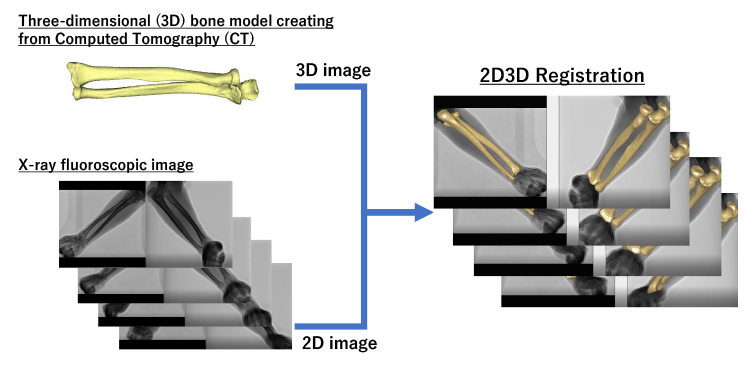
Reproduces detailed 3D motion of the upper limb
3. 3D bone morphology using statistical shape analysis
It has been previously reported that bone morphology in patients with osteoarthritis (OA) and rheumatoid arthritis (RA) differs from that of the normal knee. We have evaluated the bone morphology between OA, RA and normal knee, using statistical shape analysis. Our aim is to improve our understanding of the etiology and to develop the surgical implants.
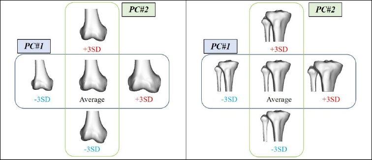
Statistical shape analysis: Femur (Left) and Tibia/Fibula (Right)
4. Application of Artificial Intelligence Technology to Orthopaedic Surgery
In collaboration with Nara Institute of Science and Technology (Prof. Sato and Assoc. Prof. Otake), we are developing new technology using artificial intelligence technology to estimate 3 dimensional (3D) bone shape from simple X-ray images without CT imaging. So far, we have constructed 3D bone shapes from simple X-ray images of normal wrist joints, and the results have been published. We are now conducting research to expand the application of this technology to other parts of the body and to disease cases such as fractures.
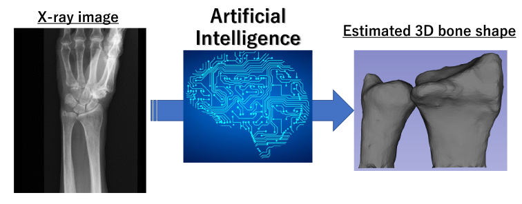
AI to estimate 3D bone shape from simple X-ray images without CT imaging
5. Bone density and stress distribution patterns on the joint
Bone deformities after fracture cause the progressive joint disorders adjacent to the deformed bone. If left untreated, osteoarthritis progresses, resulting in irreversible changes in joints. We develop the new evaluation methods to predict such joint changes in advance and use them in treatment to preserve joints before joint damage appears.
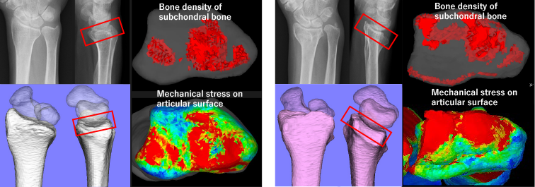
Bone density and stress distribution patterns of Normal wrist (Left) and malunited distal radius fracture (Right)
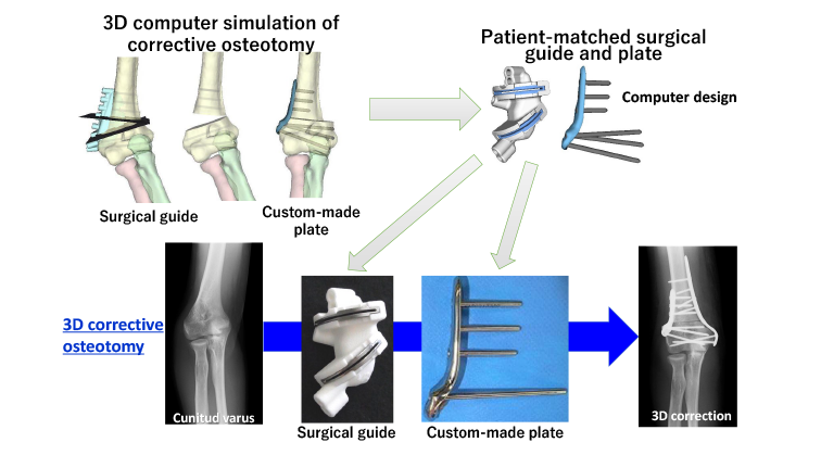
6. Development of 3D computer simulation and patient-matched surgical guide and plate for bone deformity
3D corrective osteotomy is performed using 3D computer simulation technology and patient-matched surgical guide and implant.